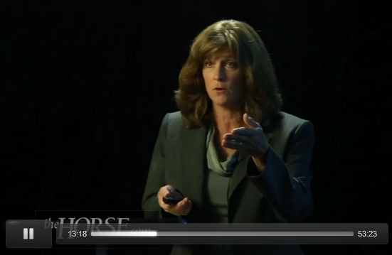
Vet examines why some laminitic feet return to soundness
Some horses recovering from laminitis and coffin bone rotation become sound even though the hoof wall no longer is parallel to the bone. Dr. Debra Taylor, DVM, looks at possible explanations for this occurrence in a video posted on thehorse.com in September 2014.
Taylor is an equine podiatrist at Auburn University in Alabama.
In Taylor’s 53-minute presentation, she discusses her journey in trying to come up with basic definitions for what constitutes a healthy equine foot and an appropriate physical exam of the foot.
Much of her presentation focuses on the back half of the foot and its soft tissue structures, more suited for absorbing concussion than the front half of the foot.
Taylor said she used to focus on the front of the hoof with laminitic horses, trying to regenerate normal parallelism between the hoof wall and coffin bone. When some of the horses became sound without parallelism, she asked herself: “What in the world are these horses walking on?”
One thing she saw was that increased heel volume seemed to play a role in a horse returning to soundness despite incomplete resolution of rotation. She hypothesized that increased heel volume might compensate for coffin bone remodeling or damage to the laminae.
She said hoof loading creates shock waves that are strong enough to crack a bone, but generally they don’t. They are absorbed through soft tissue. The front part of the foot has less soft tissue and is more rigid, making it more susceptible to shock waves. Taylor said a large frog and prominent heels are likely to play a significant role in cushioning hoof impact.
She credits farrier Pete Ramey with inspiring her to investigate the roles of the front and back of the foot. Ramey once commented that the coffin bone supports the front half of the foot and the digital cushion and lateral cartilages support the back half. She set about quantifying that ratio.
Taylor shows data on three feet comparing the size of the coffin bone in each to the size of the soft tissue structures. In the foot considered the healthiest, the lateral cartilages and digital cushion make up 159 percent of the area of the coffin bone. In the weakest foot, the soft tissue makes up less than 100 percent of the area of the coffin bone. Taylor said there’s a huge variation in nature and perhaps this type of comparison in a hoof can be useful in predicting whether the hoof is capable of taking care of that horse.
Taylor starts her presentation by talking about how horses’ feet are smart, changing in response to external factors. She wonders if the use of physical stimulation and physical therapy can create, or forge, the tissue needed for a healthy equine foot.
Working on grants, she realized there was no consensus on what a healthy foot looks like. Using several papers, she came up with the following description of a healthy foot. She provides 3-D models in her lecture to help illustrate.
Healthy hoof ratio
The foot should have more weight-bearing surface in the back half than the front using the widest part of the foot as a reference line.
Taylor does not review how to find the widest part of the foot. Some hoof experts suggest doubling the length of the central sulcus, or frog dimple, to reach the true apex of the frog, then measuring an inch back toward the heel to indicate the widest part of the foot. Farrier Gene Ovnicek provides an often-used hoof mapping protocol on his website.
Taylor said the front of the foot should be 40 to 50 percent of the weight-bearing surface of the foot and the back part of the hoof should be 50 to 60 percent of the weight-bearing surface of the foot.
Farriers who use this ratio often comment that most horses are trimmed with 60 to 70 percent of the weight-bearing surface out front of the widest part of the hoof, leading to too much pressure on the toe, prying apart of the laminae and painful abscessing.
Healthy frog dimensions
Taylor said the width of a frog should be 50 to 60 percent of its length. The depth should reach the ground, and there should not be a huge air cavity under the frog. The central sulcus, or dimple in the frog, should be wide and shallow without thrush. She shows an example of a heel that became healthier after she treated the frog for bacteria and fungus over a couple of months in muddy weather.
Healthy digital cushion
The digital cushion (a flexible layer of tissue that sits between the lateral cartilages and above the frog and cushions the back half of the foot), should be about 2 inches thick at the back of the foot and three to four fingers wide. She said veterinarians need instrumentation to evaluate the density of the digital cushion, perhaps a tool that would be similar to an A-shore scale measurement tool used to evaluate the density of rubber. She said the density of a digital cushion in decent feet is similar to that of a tennis ball or well-done steak. As for deformability, the digital cushion should have minimum deformability with maximum thumb pressure. She demonstrates how to palpate this tissue at the back of the foot. But she also said that the shape of the digital cushion as it tapers off toward front of the hoof may actually determine how effective it is in supporting the foot.
Healthy collateral cartilages
The collateral cartilages on either side of the digital cushion should be thick but slightly bendable with moderate thumb pressure and about 3 to 4 inches apart.
Healthy bars
The bars should end mid-frog and stand fairly erect. Taylor says to remove folded-over bars that may bruise the sole.
Coronary band circumference
Taylor says her team is finding that coronary band circumference is a good predictor of the volume of the internal structures. She did not provide more detail on that.
Healthy collateral grooves
She said her team was working on a paper on using the collateral grooves (the grooves alongside the frog) to predict coffin bone position. She said her team believed they would be able to use the slope of the collateral grooves to predict sole depth, coffin bone suspension off the ground, palmar angle, and maybe even heel development.
Ramey, the farrier, has done a lot of work in this area and describes the role of the collateral grooves as visual guides. On his website, he says the sole grows down from the bottom of the coffin bone. The sole corium, a bloody layer between the coffin bone and sole, is consistently a half inch away from the collateral grooves at their deepest part, no matter whether the rest of the sole is too thick or too thin. Thus, if a horse has a lot of sole, the collateral grooves will be deep. If a horse has little sole thickness, the collateral grooves will be shallow.
In Taylor’s lecture, she said the grooves gradually get deeper toward the back of the foot. The front hooves get deeper more quickly than the back feet. The collateral groove slope predicts palmar angle (the slope of the bottom of the coffin bone in the front feet). The palmar angle in the front feet averages about 6 degrees, while the angle (called the plantar angle) in the back feet is about 2 degrees.
She said an undulating, or wavy, collateral groove would be akin to a dropped arch in a human foot and cause discomfort, as would collateral grooves deeper at the frog apex, which would indicate a negative slope of the bottom of the coffin bone and warrant radiographs.
Taylor said horse feet can have pathology, even though a horse is sound, much like people can function normally even though they are on the verge of a heart attack. She thinks a lot of horses are in this state, on the verge of a hoof attack, and no one recognizes that there’s pathology in the foot.
She said she saw so many feet with pathology in her early experience that she believed such feet were normal until she learned more about the function of the foot.
In a nod to the barefoot movement, she said barefoot farriers are running ahead of science in their hypothesis that the back half of the foot is essential for overall hoof health and the back half is very smart, or adaptive.
She also quotes an ancient Greek horse expert in his day who said smooth surfaces lead to the detriment of the equine foot and horses should be stabled on cobblestone.
Taylor shows an equine foot that developed a healthy concave sole after walking on pea gravel. She said horses get addicted to shoes and hoof boots, and lack of necessary stimulation may hinder healthy development and healing of those feet.
